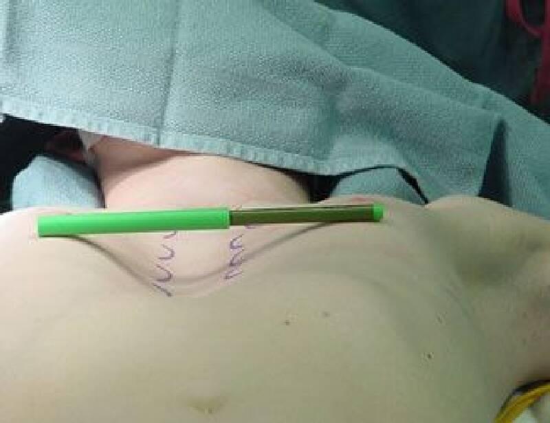
Anterior chest wall deformities are roughly examined under two main headings: •Shoemaker’s chest (Anterior chest wall is collapsed inwards.) •Pigeon chest (Anterior chest wall is protruding outwards.)

Chest Wall Deformities (Funnel Chest, Pigeon Chest)
Chest wall deformities are roughly categorized under two main headings.
Funnel chest (Chest wall is inwardly concave.)
Pigeon chest (Chest wall is outwardly convex.)
Funnel chest (Pectus excavatum) and pigeon chest (Pectus carinatum) are the most common hereditary chest wall deformities. They are observed in the community with a frequency of 0.01-0.1%.
They are most commonly encountered in the western Black Sea region of our country.
In the deformity of the funnel chest, exercise restriction and shortness of breath may occur due to a decrease in the volume of the chest cage. In some patients, leaks in the heart valves may occur due to the displacement of the heart. These findings are not observed in pigeon chest deformity.
The exact causes of chest wall deformities are not fully understood. Genetic factors are also considered among the possible etiological factors. 35% of patients have a family history of chest wall deformity.
Deformity can be evident from birth or may occur within a year after birth and tends to progress. It usually stabilizes around the ages of 7-9. In most cases, the depression is in the sternal body, and its depth is maximum at the xiphoid level. The xiphoid prominence may be separate, rotated, or shifted to one side in some cases.
Funnel chest (Pectus excavatum):
It is the most common chest wall deformity. There is an inward concavity in the sternum (chest wall bone) and ribs. The participation of the first two ribs in the concavity is rare. The deformity can be centralized or asymmetrical. In asymmetrical cases, the depression is more often seen on the right side, and there is a rotation to the right around the long axis of the sternum. In girls with this type of deformity, the right breast may be smaller than the other. The frequency of Pectus excavatum deformity is one in 300 births in the general population. The male-to-female ratio is 3:1. Symptoms related to the disease may appear in childhood and are related to the degree of deformity; mild deformities are generally asymptomatic. With more advanced deformities, chest pain, palpitations, and restricted movement may occur with effort. Fatigue is the most common symptom. Mitral valve prolapse may be the cause of symptoms in these cases and is seen in 15% of cases.
Children with Pectus excavatum are generally thin and flat-chested, with a slight hump. Scoliosis is present in a quarter of cases. In patients with severe funnel chest deformities, the heart is often displaced to the left.
Pigeon chest (Pectus carinatum):
Pectus carinatum deformity is an excessive outward projection of the chest wall. It is seen less frequently (one-fifth) than Pectus excavatum deformity. The most common form is the symmetrical protrusion of the sternum body with lower cartilage ribs. It is three times more common in males than females. It usually occurs in childhood and adolescence. Familial transmission is present in a quarter of patients with pigeon chest. Scoliosis accompanies the condition in one-fifth of patients.
In pigeon chest deformity, the chest wall bone and, consequently, the rib cartilages are most affected. Sometimes, mixed-type deformities may also occur. Usually, the deformity is symmetrical, but in some cases, it may be asymmetrical.
Chest wall deformities do not restrict the normal lifespan of individuals. Psychological discomfort may be present in these individuals. Funnel chest and pigeon chest can be successfully corrected through surgery for cardio-pulmonary, psychological, and cosmetic reasons. Various techniques have been used to correct these deformities.
The treatment of chest wall deformities was first performed in 1911 by Meyer and in 1913 by Sauerbruch. In 1949, Ravitch used his developed method. A modified version of this open surgical method is still used today.
In this method, the sheaths covering the ribs and cartilages participating in the deformity are removed while preserving their integrity. After correcting the deformed cartilages, they are repositioned and sutured. One or two wedge incisions are made in the flat bone of the chest wall that is primarily affected by the deformity. After correction, it is fixed with Titanium plate screws or absorbable plate screws. If the cartilage at the lower end of the chest wall bone is deformed, it is also corrected. After all these procedures, the bone and muscle layers are closed in their original form, and the process is completed. The best part of this method is that the surgery is completed in one session. There is no need to remove the used plates and screws. The patient continues their life comfortably in the postoperative period.
Recently, in our country, there is a method developed by Donald Nuss. This method is used for funnel chest.
In this method, the procedure is performed endoscopically. It is done in a shorter time than the previous method. In this method, the basic procedure is the rotation and elevation of the bar by steel bar to lift the area with the deformity (depression), and fixing the steel bar to the chest wall. This bar must be removed 2-4 years later. For this, a new operation is required. Sometimes this waiting period can exceed 4 years. Collapsed lung, lung tear and bleeding, and more importantly, life-threatening tears in the heart muscle can occur rarely.
In our hospital, the basis of the treatment of chest wall deformities is the surgical correction of the deformed part. We achieve satisfactory results from our patients.
Titanium or absorbable plates and screws are used in surgery.
Titanium plates are permanent and do not need to be removed. For this reason, there is no need for a second operation. Absorbable plates are absorbed by the body within 6 months. The average length of hospital stay is 5 days.
Images before and after funnel chest surgery.
Images before and after pigeon chest surgery.


